What Is Tongue Cancer?
Tongue cancer is a type of oral cancer that develops in the tissues of the tongue. Tongue cancer occurs in the front two-thirds of the tongue within the oral cavity or in deeper tissues closer to the throat, depending on the tumor location. It develops when abnormal cells grow out of control, and the most common form is tongue squamous cell carcinoma, which begins in the thin cells lining the surface of the tongue.
A carcinoma of oral tongue may appear as a sore, lump, ulcer, thickened area, red patch, white patch, or tissue change that does not heal. Some lesions are painful, while others may cause little discomfort in the early stages. This is why professional evaluation is essential when a tongue abnormality lasts longer than two weeks.
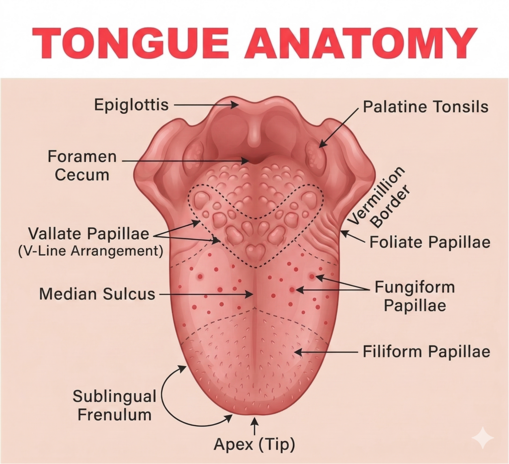
Certain tumors may develop along the side of the tongue. Lateral border tongue cancer is especially important to evaluate because this area is a common location for suspicious oral lesions. Other cases may involve the base of the tongue, which is anatomically distinct from oropharyngeal cancer and may require coordinated evaluation with head and neck oncology specialists.
At Love Your Jaws, we evaluate tongue changes carefully and recommend biopsy or additional imaging when needed.
What Are the Early Signs and Symptoms of Tongue Cancer?
Tongue cancer symptoms are among the common symptoms that can appear subtly at first. A small sore, unusual patch, or firm area may seem minor at first but should not be ignored if it persists. The most important warning sign is a lesion that does not heal, and oral cancer on the tongue often presents as a persistent, non-healing sore.
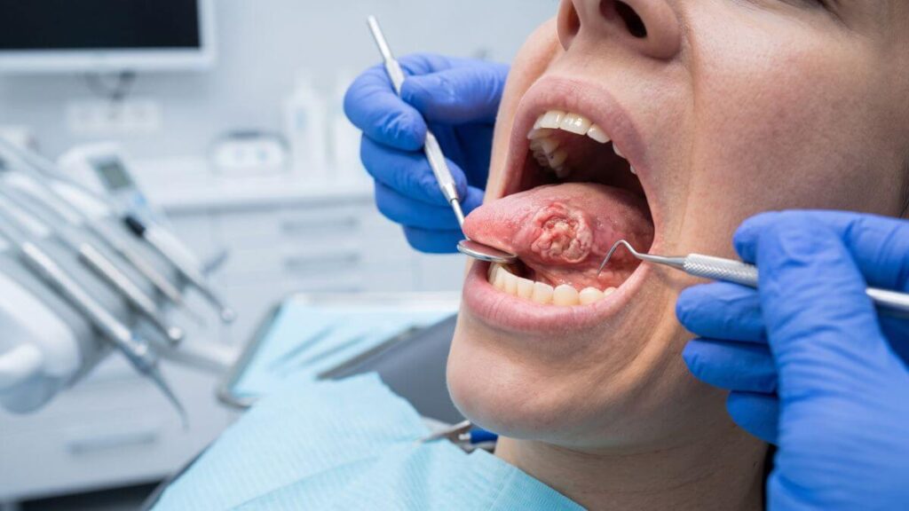
Common early signs of tongue cancer may include a persistent ulcer, tongue pain, unexplained bleeding, numbness, difficulty moving the tongue, pain with swallowing, a white or red patch, or an unexplained lump that feels firm or irregular. A sore on the side of the tongue not healing should always be examined by an experienced oral surgeon.
Some patients notice a tongue lesion white patch, while others see a red spot on tongue cancer concern that looks different from surrounding tissue. A painful or painless exophytic tongue mass may grow outward from the surface, while an endophytic tongue tumor may grow deeper into the tissue and feel firm beneath the surface.
Other symptoms may include a painful lump on tongue oncologist concern, difficulty speaking, pain radiating toward the ear, or dysphagia tongue cancer symptom, meaning difficulty swallowing. In some cases, a persistent ulcer may represent tongue ulcer malignant transformation, which requires prompt evaluation and possible biopsy.
Patients searching for Tongue lesion evaluation Miami Beach or a Miami emergency tongue biopsy clinic often come to Love Your Jaws because they want fast answers from a trusted oral surgery team.
Key Differences: Oral vs. Base of Tongue
| Feature | Oral Tongue Cancer (Front) | Base of Tongue Cancer (Back) |
| Visibility | Easily seen during a mirror exam. | Often hidden; requires specialized scopes. |
| Primary Risk | Tobacco and heavy alcohol use. | Often linked to the HPV-16 virus. |
| First Signs | Visible sores or white/red patches. | Difficulty swallowing or a lump in the neck. |
| Diagnosis | Simple in-office biopsy. | May require fiber-optic endoscopy. |
How Is Tongue Cancer Diagnosed?
Tongue cancer diagnosis begins with a detailed oral examination, and when a lesion appears suspicious or deeper spread is a concern, imaging tests may also be recommended. We evaluate the lesion’s size, color, texture, borders, firmness, depth, location, and whether it bleeds or causes pain. We also assess tongue movement, surrounding tissues, lymph nodes, and any symptoms involving swallowing or speech.
People who visit a dentist regularly may have suspicious changes noticed earlier during oral cancer screenings.
When a lesion appears suspicious, a tongue cancer biopsy may be recommended. During this procedure, a small tissue sample is removed and sent to a pathology laboratory for microscopic evaluation to check for cancerous cells. Patients searching for a tongue biopsy doctor in Miami FL often need this step to determine whether the lesion is benign, precancerous, or malignant.
The tongue biopsy pathology report provides the diagnosis and helps determine the next step. If cancer is confirmed, further testing may be needed to evaluate tumor depth, spread, and lymph node involvement. tongue cancer staging TNM helps classify the tumor based on size, nodal involvement, and whether cancer has spread beyond the original site.
In advanced cases, imaging may evaluate deeper structures, including concern for deep lingual artery tumor invasion or metastatic tongue cancer lymph nodes. Patients may also be referred to a head and neck oncology tongue specialist or head and neck surgeon for tongue tumor in Miami when multidisciplinary treatment is needed.
Patients comparing advanced cancer resources such as Sylvester Cancer Center tongue oncology or University of Miami tongue cancer clinic may still begin with expert oral biopsy and surgical evaluation at Love Your Jaws.
Clinical Palpation: Manually feeling the tongue and neck to check for lumps or “indurated” (hardened) tissues.
Expert Biopsy: A small tissue sample is removed under local anesthesia and sent to an oral pathologist. This is the only definitive way to confirm a diagnosis.
3D Imaging: We utilize 3D Cone Beam CT (CBCT) and, if necessary, coordinate MRI or PET scans to determine if the cancer has affected the jawbone or spread to the lymph nodes in the neck.
Who Is at Risk for Developing Tongue Cancer?
Tongue cancer can affect many different types of patients, and there are several risk factors that increase the likelihood of developing the disease. Primary causes of oral cancer include tobacco use, heavy alcohol consumption, and HPV infection, while smokeless tobacco, chronic irritation, immune suppression, poor oral hygiene, previous oral lesions, and other factors may also increase risk. A poor diet may increase the risk of developing tongue cancer, while a healthy diet rich in fruits and vegetables may help lower it.
Some tongue cancers are associated with viral factors, including HPV positive tongue cancer, which may behave differently from tobacco-related cancers and may require specialized evaluation. Patients with long-standing white or red tongue lesions should also seek professional assessment, especially if the area changes in size, shape, color, or texture.
Age may increase risk, and it is more common in males, but younger patients can also develop tongue cancer. This is why the incidence is increasing among younger non-smokers, including some young adults, and any suspicious lesion should be evaluated based on clinical behavior rather than age alone.
At Love Your Jaws, we review personal risk factors, lesion history, oral habits, medical background, and symptoms before recommending the next diagnostic step. Early evaluation gives patients the best opportunity for timely diagnosis and treatment.
What Treatment Options Are Available for Tongue Cancer?
Treatment depends on the tumor’s size, depth, location, stage, pathology, and whether lymph nodes are involved. At Love Your Jaws, we help patients understand the diagnosis and coordinate the right treatment pathway with the appropriate specialists when needed. Treatment for oral cancer may involve surgery, radiation therapy, chemotherapy, or a combination depending on the stage and spread.
For early localized tumors, partial glossectomy surgery may be recommended to remove the affected portion of the tongue while preserving as much function as possible. More advanced tumors may require tongue tumor resection, and in rare extensive cases, total glossectomy reconstruction may be considered as part of complex cancer care.
If cancer has spread to lymph nodes or carries a high risk of spread, neck dissection tongue cancer may be recommended by the oncology surgical team. Additional treatment may include radiation therapy for tongue cancer or chemotherapy for squamous cell carcinoma tongue, depending on stage and pathology. Radiation therapy uses high doses of radiation to kill cancer cells, and broader systemic therapies may also be used when disease is advanced.
When larger areas of tissue are removed, free flap reconstruction tongue surgery and reconstructive surgery may help restore structure, speech, and swallowing after tumor removal. Patients may also need speech therapy after glossectomy to improve communication, swallowing, and quality of life following surgery.
Patients searching for a tongue cancer surgeon in Miami, glossectomy surgery specialist in Miami, or oral oncologist for tongue cancer in South Florida tongue cancer can trust Love Your Jaws for careful evaluation, biopsy, surgical guidance, and coordinated care planning.
What Is Recovery Like After Tongue Cancer Treatment?
Recovery after tongue cancer treatment depends on the size and location of the tumor, the type of surgery performed, whether reconstruction was required, and if additional therapies such as radiation or chemotherapy are included in the treatment plan. Every patient heals differently, which is why we create individualized recovery plans focused on restoring oral function while supporting long-term health.
Immediately after surgery, patients may experience swelling, discomfort, temporary changes in speech, difficulty swallowing, and dietary restrictions. Most patients begin with a modified or soft-food diet before gradually returning to more regular foods as healing progresses. Our team carefully monitors every stage of recovery to ensure proper wound healing and minimize complications.
Patients who undergo more extensive tongue surgery may benefit from speech therapy, swallowing rehabilitation, nutritional counseling, and coordinated follow-up with oncology specialists. We work closely with every patient to restore communication, eating function, and overall quality of life as efficiently as possible.
What Is the Outlook for Patients with Tongue Cancer?
The outlook for tongue cancer depends on several important factors, including how early the cancer is diagnosed, the size and depth of the tumor, lymph node involvement, tumor biology, and the patient’s overall health; as part of head and neck cancer, oral and oropharyngeal cancers account for about 85% of cases, according to the National Cancer Institute.
Patients diagnosed with stage 1 tongue cancer symptoms often have significantly more treatment options because the disease is detected before becoming more advanced. This highlights the importance of evaluating persistent tongue ulcers, white or red patches, and unexplained tissue changes as early as possible.
While many patients search online for the tongue cancer survival rate, survival statistics show the five-year survival rate for tongue cancer is approximately 57%, though an individual’s outlook still varies based on the specific diagnosis, pathology findings, response to treatment, and long-term follow-up. Early detection remains the most important factor influencing successful treatment and long-term disease control, since delayed diagnosis contributes to a high death rate, and tongue cancer death rates have been reported as higher than cervical cancer rates.
At Love Your Jaws, we focus on identifying suspicious lesions early, providing timely biopsy procedures, and coordinating comprehensive treatment that gives every patient the best possible opportunity for a positive outcome.
How Much Does Tongue Cancer Treatment Cost in Miami?
The cost of tongue cancer treatment varies depending on the complexity of the diagnosis, imaging studies, biopsy procedures, pathology analysis, surgical treatment, hospital services, anesthesia, reconstructive procedures, and any additional therapies recommended after surgery.
Some patients require only biopsy and removal of a small localized lesion, while others may need more advanced surgery, neck procedures, reconstructive treatment, radiation therapy, chemotherapy, or multidisciplinary oncology care.
During your consultation, we explain every recommended diagnostic and treatment step before care begins. Our team provides transparent information regarding anticipated costs, insurance considerations when applicable, and the treatment sequence best suited to your diagnosis.
Our priority is helping every patient fully understand both their condition and their available treatment options before making any decisions.
Which Miami Areas Do We Serve for Tongue Cancer Treatment?
Love Your Jaws proudly provides comprehensive tongue cancer evaluation and oral surgery services for patients throughout Miami and South Florida from one modern, centrally located oral surgery center. While we practice from a single location, patients travel from surrounding communities because they value experienced surgical care, advanced diagnostics, and personalized treatment planning.
Oral Cancer Tongue Treatment in South Miami
Patients from South Miami trust our team for prompt evaluation of persistent tongue lesions, biopsy procedures, surgical consultations, and individualized oral cancer treatment planning.
Oral Cancer Tongue Treatment in Coral Gables
Residents of Coral Gables choose Love Your Jaws for advanced tongue cancer evaluations, expert biopsy services, and coordinated care designed to support early diagnosis and successful treatment.
Oral Cancer Tongue Treatment in Brickell
Many patients from Brickell visit our office for comprehensive assessment of suspicious tongue abnormalities, second opinions, and personalized oral surgery consultations.
Oral Cancer Tongue Treatment in Key Largo
Patients from Key Largo frequently travel to our Miami surgical center for expert tongue lesion evaluation, biopsy procedures, and coordinated oral cancer care delivered by an experienced oral surgery team.
Oral Cancer Tongue Treatment in Pinecrest
Residents of Pinecrest rely on Love Your Jaws for early diagnosis of suspicious tongue lesions, comprehensive surgical evaluations, and compassionate patient-centered treatment planning.
Oral Cancer Tongue Treatment in Fisher Island
Patients from Fisher Island appreciate our modern diagnostic technology, meticulous clinical evaluations, and individualized treatment recommendations for tongue cancer and other oral pathology conditions.
Oral Cancer Tongue Treatment in Coconut Grove
Many Coconut Grove residents choose our practice for timely biopsy procedures, careful evaluation of persistent tongue ulcers, and coordinated oral oncology consultations focused on preserving long-term oral health.
Why Choose Love Your Jaws for Oral Cancer Tongue Treatment in Miami?
When a suspicious tongue lesion is discovered, experience and timely diagnosis matter. At Love Your Jaws, we combine advanced oral pathology expertise, meticulous surgical techniques, and compassionate patient care to provide comprehensive tongue cancer evaluation and treatment planning.
Close to 58,500 Americans will be diagnosed with oral cancer this year.
Dr. Kroum Dimitrov is recognized by many patients as one of the leading oral surgeons in Miami, providing expert assessment of tongue lesions, advanced biopsy procedures, and personalized surgical care for patients with suspected or confirmed oral cancer. Every patient receives a thorough examination, clear communication, and an individualized treatment strategy based on the latest evidence-based oral and maxillofacial surgery protocols.
Our practice emphasizes early detection, minimally invasive biopsy techniques whenever appropriate, advanced digital imaging, and close collaboration with head and neck oncology specialists whenever multidisciplinary treatment is required, including coordination of therapies that engage the immune system when appropriate. Whether you require diagnostic evaluation, surgical management, or coordinated oncology care, our goal is to preserve oral function, speech, swallowing, and quality of life while achieving the best possible clinical outcome.
Patients searching for specialized tongue cancer care throughout Miami choose Love Your Jaws because we deliver comprehensive oral surgery services with precision, transparency, and compassion. From your first consultation through diagnosis, treatment, recovery, and long-term follow-up, we are committed to providing exceptional care tailored to your individual needs.
If you notice a persistent sore, lump, ulcer, white patch, red patch, or any other unusual change on your tongue that has not healed within two weeks, we encourage you to schedule an evaluation promptly. Early diagnosis remains one of the most powerful tools for improving treatment options and protecting your long-term health.
Why Choose Dr. Dimitrov
Elite Training: Earned his DMD from the University of Connecticut and completed residency at the University of Miami/Jackson Memorial Hospital, where he served as Chief Resident.
Proven Leadership: Awarded the prestigious Professor’s Cup for excellence in surgery, patient care, and academic achievement.
Meet Dr.Kroum Dimitrov