

Cone Beam CT Scan: 3D Imaging That Changes How We See Your Smile
When it comes to diagnosing oral and maxillofacial conditions, a flat 2D X-ray can only tell part of the story. Bone structure, nerve pathways, sinus cavities, and the precise angle of a tooth root all exist in three dimensions – and that’s exactly how we believe they should be seen. 3D imaging captures the depth, shape, and volume of oral structures, allowing us to create digital 3D models for comprehensive visualization and more precise treatment planning.
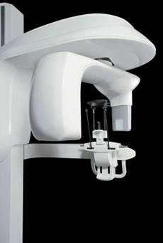
That’s why Love Your Jaws Surgery Center, Miami’s first robotic dental implant solution, uses the i-CAT FLX – a state-of-the-art 3D imaging platform developed as an advanced tool for dentistry. This technology enables detailed visualization of the patient’s anatomy, supporting more accurate diagnoses and treatment planning by providing a comprehensive representation of anatomical structures.
The result? 3D imaging has revolutionized medical diagnosis and treatment since the 1990s by providing healthcare professionals with deeper insights into complex oral and maxillofacial conditions. More accurate diagnoses, smarter treatment planning, fewer surprises, and care delivered with confidence – all with significantly less radiation than a conventional medical CT scan.
What Is a Cone Beam CT Scan?
A cone beam CT scan – often called a CBCT scan or 3D dental CT scan – is a specialized type of x-ray imaging that uses a cone-shaped x-ray source to create detailed, three-dimensional images of your teeth, jawbones, sinuses, and surrounding soft tissues in the head and neck.
During the imaging process, the x-ray source and detector are mounted on a rotating gantry that moves around your head, capturing multiple images from different angles. These images are acquired using ionizing radiation, but CBCT delivers a lower dose compared to traditional CT scans. The detector collects the data needed for 3D reconstruction, and the entire process is quick and comfortable, with you standing upright – no narrow tunnels or lying down required.
The reconstruction process then creates a single, comprehensive 3D image from the multiple images captured during the scan, allowing for highly detailed digital representations of your anatomy. A typical cone beam CT procedure takes between 20 to 40 seconds for a complete volume scan.
What we get back is a digital map of your anatomy so detailed it’s measured in micrometers (the i-CAT FLX captures detail down to 90 microns – roughly the width of a human hair).
How the i-CAT FLX Cone Beam CT Works
1. A Quick, Comfortable Upright Scan
During the thorough examination, you stand or sit upright in the open, face-to-face design of the scanner, which prioritizes patient safety by being non-invasive and comfortable. There’s no enclosed tube, no need to lie back, and no contrast injection. Patients of all sizes – including those in wheelchairs – can be comfortably accommodated. The use of advanced CBCT technology in this examination reduces radiation exposure compared to traditional imaging methods, making it a safer option for patients undergoing dental procedures.
2. A Single Rotation Captures Everything
The imaging process involves the x-ray source rotating around your head, capturing multiple images from different angles for detailed 3D reconstruction. The scanner completes a full rotation, acquiring hundreds of high-resolution images in about 20–30 seconds. Typically, a cone beam CT procedure takes between 20 to 40 seconds for a complete volume scan.
3. Powerful Software Builds Your 3D Model
Specialized software performs reconstruction of the images, building a true 3D digital model of your mouth, jaw, and skull – viewable from any angle, any depth, and any cross-section. After reconstruction, doctors and radiologists conduct a detailed analysis of the volumetric data from MRI, CT, or 3D ultrasound scanners, allowing them to visualize complex internal anatomy and prepare for highly precise surgery. Dr. Dimitrov can rotate, zoom, and “slice through” the model to see exactly what’s happening at any point in your anatomy.
4. Diagnosis and Treatment Planning
The 3D model undergoes detailed analysis to support accurate diagnosis and treatment planning. Whether you’re being evaluated for dental implants, wisdom teeth removal, jaw surgery, or a complex pathology, having a true 3D view means Dr. Dimitrov plans your procedure before any instrument touches you. The results of this analysis are also shared with your referring physician to ensure collaborative care and optimal patient management. Using cone beam computed tomography (CBCT) provides detailed 3D images that enhance diagnostic accuracy and treatment outcomes, while also reducing radiation exposure compared to traditional imaging methods – helping to alleviate patient anxiety and improve your overall experience.
A Lower-Dose Approach: The ALARA Principle
One of the most important advantages of our i-CAT FLX cone beam CT system is its commitment to the ALARA principle – “As Low As Reasonably Achievable.” CBCT uses ionizing radiation to acquire high-quality 3D images, but is specifically designed to maximize patient safety by minimizing exposure. In fact, compared to traditional dental CT, CBCT significantly reduces radiation exposure while still providing detailed 3D images, making it a safer option for patients who require advanced imaging for diagnosis and surgical planning.
Here’s how the i-CAT FLX makes that possible:
Adjustable, collimated fields of view. The i-CAT FLX allows Dr. Dimitrov to scale and collimate the scan height to capture only the area of interest – from a focused single-arch view for an implant case all the way up to a full craniofacial scan when needed. Smaller fields of view mean lower radiation exposure, because we are scanning only the region your treatment actually requires.
Significantly lower dose than medical CT. Cone beam CT scans deliver only a small fraction of the radiation exposure of a conventional medical (hospital) CT scan, while still providing comparable – and in some cases superior – diagnostic detail for dental and maxillofacial applications. Peer-reviewed research has measured the i-CAT FLX’s effective dose at roughly two orders of magnitude less than a typical medical CT head scan. This reduction in ionizing radiation exposure is a key factor in enhancing patient safety.
QuickScan and QuickScan+ low-dose protocols. For appropriate cases, the i-CAT FLX’s QuickScan+ mode can capture a full 3D scan at a radiation dose comparable to – or even less than – a standard 2D panoramic X-ray. This is especially valuable for follow-up imaging and cases where ultra-high resolution isn’t required. (Image quality is proportional to dose, so Dr. Dimitrov selects the appropriate protocol for each individual diagnostic need.)
Faster scan times mean less exposure. A standard i-CAT FLX scan completes in just a few seconds, and QuickScan+ protocols can capture a full 3D image in as little as two seconds – dramatically reducing the time you’re exposed to imaging radiation compared to slower scanners.
Seated positioning for fewer retakes. Unlike many cone beam CT systems that require you to stand, the i-CAT FLX features a comfortable seated design with an Ergonomic Stability System (ESS) built specifically to minimize patient movement. Less movement means fewer retakes – and fewer retakes mean less cumulative radiation exposure.
Protocol selection tailored to every patient. Dr. Dimitrov adjusts field of view, voxel size, and exposure protocol to match the specific clinical need – whether that’s a small implant site, a TMJ evaluation, an airway study, or a complete maxillofacial scan. The smallest appropriate scan is always the right scan.
Less radiation. More information. That’s a win in every direction.
Why Cone Beam CT Matters for Your Care
Adding 3D imaging to our practice isn’t a luxury – it’s a meaningful upgrade to nearly every type of procedure Dr. Dimitrov performs.
More accurate diagnoses. Doctors use detailed analysis and advanced visualization of 3D images, such as those from cone beam computed tomography (CBCT), to enhance diagnostic accuracy and treatment planning. Hidden infections, cracked roots, impacted teeth, cysts, tumors, and nerve proximity issues that are invisible on 2D X-rays are clearly visible in 3D.
Safer, more precise oral surgery. The comprehensive visualization provided by 3D imaging technologies like CBCT allows for a better evaluation of complex oral and maxillofacial structures, improving surgical outcomes and reducing the risk of complications by knowing the exact location of nerves, sinuses, and bone density before surgery.
Perfect partner for robotic implant surgery. As Miami’s first robotic dental implant practice, we rely on detailed 3D scans to plan implants with our YOMI robot. The CBCT scan is the digital blueprint the robot uses to place every implant with sub-millimeter precision.
Fewer appointments, faster treatment. Because we can perform a wider range of diagnoses in-office, you don’t have to travel to a separate imaging center or wait days for results.
Better patient education. Dr. Dimitrov can show you exactly what’s happening in your own mouth – on screen, in 3D – and walk you through your treatment options with clarity you simply can’t get from a 2D film.
Seamless collaboration with your referring doctor. Your 3D images can be securely shared with your referring dentist, orthodontist, or specialist, allowing your full care team to collaborate on the best plan for you. 3D imaging has transformed dentistry by providing more detailed images than traditional 2D X-rays, setting a new standard for patient care and interdisciplinary teamwork.
What Can a Cone Beam CT Scan Be Used For?
The i-CAT FLX supports an exceptionally broad range of oral and maxillofacial applications, enabling detailed examination and analysis for a wide variety of dental and maxillofacial conditions. These advanced CBCT scans allow doctors and radiologists to interpret high-quality 3D images for precise diagnosis and treatment planning. Applications include:
- Dental implant planning and placement, including full-arch and robotic-guided cases, with doctors and radiologists analyzing the scans to guide optimal outcomes
- Wisdom teeth and impacted tooth evaluation, with precise nerve mapping and thorough examination by doctors for safe extraction
- Bone grafting and sinus lift assessment, using 3D imaging analysis to evaluate bone quality and volume
- TMJ (temporomandibular joint) evaluation, where doctors rely on detailed imaging for accurate diagnosis
- Orthodontic and orthognathic surgical planning, utilizing 3D scans to create digital models for customized treatment
- Endodontic (root canal) diagnosis – locating extra canals and fractures invisible on 2D X-rays, with radiologist review for comprehensive analysis
- Pathology and tumor assessment, with doctors and radiologists collaborating on image interpretation
- Airway and sleep apnea evaluation, using 3D analysis to assess airway space and structure
- Trauma assessment of the jaw, teeth, and facial structures, with detailed examination supporting precise care
- Sinus and ENT-related imaging, for comprehensive evaluation of nasal cavity and sinuses
- 3D imaging scans can also be used to create customized, perfectly fitting prosthetics, enhancing patient outcomes through tailored solutions.
Full Mouth and Facial Imaging: Beyond Just Your Smile
When it comes to truly understanding your oral health, seeing just your teeth isn’t enough. Full mouth and facial imaging with cone beam computed tomography (CBCT) gives your dental team a comprehensive view of all your oral and maxillofacial structures – not just your smile, but your entire mouth, jaw, facial bones, and surrounding tissues. This advanced medical imaging technology uses a cone-shaped X-ray beam to capture multiple images from different angles, which are then reconstructed into a single, detailed 3D model of your anatomy.
With this technology, dentists and oral surgeons can visualize every aspect of your oral and facial structures with remarkable clarity. Whether you need dental implants, orthodontic evaluation, or complex surgical planning, CBCT provides high-quality images that reveal the relationships between teeth, bone, nerves, sinuses, and soft tissues. This comprehensive view is essential for accurate diagnosis and precise treatment planning, especially in more complex cases where traditional X-rays might miss critical details.
By leveraging the power of CBCT, your care team can detect hidden issues, plan treatments with greater confidence, and deliver care that’s tailored to your unique anatomy. The result is improved diagnostic accuracy, better patient care, and outcomes you can trust. At Love Your Jaws, we believe that seeing the whole picture, literally, leads to better, safer, and more effective dental and surgical solutions.
2D X-Ray vs. Cone Beam CT Scan
| Traditional 2D X-Ray | Cone Beam CT (i-CAT FLX) | |
|---|---|---|
| Dimension | Flat, 2D image | Full 3D model with added z-axis depth for precise measurement, spatial analysis, and advanced visualization |
| Detail level | Limited overlap, can hide problems | Micron-level precision (90 μm) |
| Anatomy visible | Teeth and surface bone | Bone, nerves, sinuses, soft tissue context; comprehensive representation of anatomical structures |
| Radiation | Very low | Low – focused to the region of concern |
| Planning value | Basic screening | Comprehensive surgical planning, enhanced diagnostic accuracy, and detailed analysis |
| Use case | Routine checkups, cavity detection | Implants, surgery, complex diagnostics |
The 3D imaging process involves systematic steps of image acquisition, reconstruction, and post-processing, resulting in detailed visualizations that support precise analysis and treatment planning. This comprehensive approach enables immersive digital experiences and robotic navigation, setting a new standard for patient care.
Cone Beam CT vs. Traditional CT Scan: What’s the Difference?
While both cone beam computed tomography (CBCT) and traditional CT scans are powerful medical imaging modalities, they are designed for different purposes and offer distinct advantages – especially when it comes to imaging the oral and maxillofacial region.
Traditional CT scans use a fan-shaped X-ray beam and are typically used for imaging larger areas of the human body, such as the chest or abdomen. In contrast, CBCT uses a cone-shaped X-ray beam that rotates around the head, capturing hundreds of images from different angles in a single scan. This approach allows CBCT to produce highly detailed, three-dimensional images of oral and maxillofacial structures, including bone, teeth, soft tissues, and the nasal cavity.
One of the most significant benefits of CBCT is its lower radiation exposure compared to traditional CT scans. This makes CBCT a safer choice for patients who may require multiple scans over time. Additionally, the image quality of CBCT is optimized for dental and facial anatomy, providing higher resolution and better contrast for evaluating bone structure, soft tissues, and complex anatomical relationships. This level of detail is crucial for precise treatment planning in dental implants, jaw surgery, and other specialized procedures.
In summary, while both imaging technologies have their place in medical diagnosis, CBCT stands out for its ability to deliver high-quality, detailed images with less radiation – making it the preferred choice for dental and maxillofacial imaging. At Love Your Jaws, we use CBCT to ensure every patient receives the most accurate diagnosis and the safest, most effective treatment plan possible.
What to Expect During Your CBCT Scan
If you’ve never had a cone beam CT scan before, here’s the good news: it’s a non-invasive examination designed with patient safety in mind, making it one of the easiest imaging experiences in modern medicine.
You’ll stand or sit comfortably in the open, upright scanner. You’ll be asked to bite gently on a small positioner and hold still – no different than holding still for a regular dental X-ray. The scanner rotates once around your head, taking about 20–30 seconds total. No contrast dye, no IV, no enclosed space. The process uses advanced technology to minimize radiation exposure while providing precise, detailed 3D imagery for accurate diagnosis and surgical planning. Within moments, your 3D scan is ready on Dr. Dimitrov’s monitor for review with you.
See Your Smile in 3D – At Love Your Jaws Surgery Center
At Love Your Jaws Surgery Center, we believe better imaging leads to better outcomes. Our advanced 3D imaging technology is used to create detailed digital models of your anatomy, providing enhanced visualization for both diagnosis and treatment planning. Whether you’re being evaluated for dental implants, robotic-guided implant surgery, wisdom teeth removal, or another oral surgery procedure, the i-CAT FLX cone beam CT gives Dr. Dimitrov and our team of doctors the clearest possible view of your anatomy. This technology also allows us to create customized, perfectly fitting prosthetics tailored to your needs. Doctors and referring physicians collaborate to interpret and share your imaging results, ensuring optimal patient care through seamless communication and expert insight. With these advanced tools, you can be confident that your treatment plan is built on real data, not guesswork.
If your referring dentist has recommended a 3D scan, or if you’d like to learn more about how cone beam CT imaging fits into your care, contact our Miami office to schedule a consultation. We’ll walk you through your scan, explain what we see, and answer every question you have.
FAQ:
Coverage varies by plan and the medical necessity of the scan. Our team will help you understand your benefits before your appointment.
No. There’s no sensation during a CBCT scan. You’ll simply hold still as the scanner rotates around your head once.
Yes. CBCT scans use a significantly lower radiation dose than conventional medical CT scans. The i-CAT FLX’s selectable field of view allows us to limit your exposure to only the area of concern, following the ALARA principle.
The scan itself takes 20–30 seconds. The complete appointment – including setup and reviewing the results – is typically 10–15 minutes.
Why Choose Dr. Dimitrov

Elite Training: Earned his DMD from the University of Connecticut and completed residency at the University of Miami/Jackson Memorial Hospital, where he served as Chief Resident.
Proven Leadership: Awarded the prestigious Professor’s Cup for excellence in surgery, patient care, and academic achievement.
Meet Dr.Kroum Dimitrov